
Early embryonic stage (1-5 days)
Scale: 200:1
- 1-day embryo: Fertilized egg with an external hyaline membrane. Blastocyst size: 0.2 mm
- 2-day embryo: Two-cell stage, consisting of two oval spheres with an external membrane (zona pellucida) and 2-3 polar bodies. Blastocyst size: 0.2 mm
- 3-day embryo: Morula, consisting of approximately 16 oval spheres resembling mulberries, surrounded by the zona pellucida. Blastocyst size: 0.2 mm
- 4-day embryo: Blastocyst surrounded by trophoderm, with an internal cluster of cells at one end of the cavity and a zona pellucida membrane. Blastocyst size: 0.2 mm
- 5-day embryo: Blastocyst with the zona pellucida removed. Blastocyst size: 0.25 mm
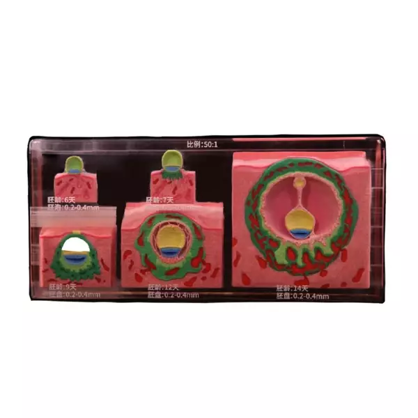
Early embryonic stage (6-14 days)
Scale: 50:1
- 6-day embryo: Beginning of implantation, attachment of the trophoblast to the endometrium. Blastocyst size: 0.25 mm
- 7-day embryo: Blastocyst partially embedded in the endometrium, trophoderm differentiates into cytotrophoderm and syncytiotrophoderm. Blastocyst size: 0.25 mm
- 9-day embryo: Blastocyst deeply penetrates the endometrium, implantation hole is closed by a coagulation plug, primary amniotic sac and yolk sac are formed. Size of embryonic disc: 0.2 mm
- 12-day embryo: Implantation completed, extraembryonic mesoderm and cavity appear, and trophodermal lacunae fill with maternal blood. Size of embryonic disc: 0.3 mm
- 14-day embryo: Secondary yolk sac forms, primary yolk sac degenerates, ectodermal vesicles form. External cavernous bodies and somites become visible, primary chorion forms. Size of embryonic disc: 0.4 mm
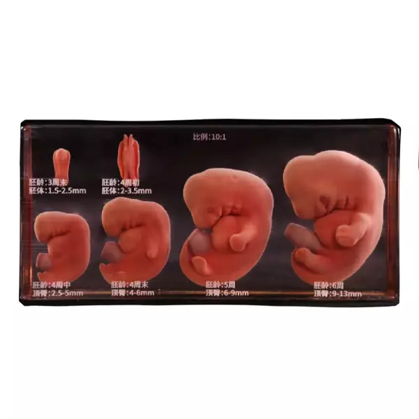
Embryonic stage (3-6 weeks)
Scale: 10:1
- End of week 3: Disc-shaped embryo evolves into a barrel-shaped form, head, tail, and lateral folds form, closure of the neural groove begins, 4-8 pairs of somites appear. Body length: 2.0 mm
- Early week 4: Barrel-shaped embryo, neural groove continues to close, anterior and posterior neural pores visible. Body segments: 8-12 pairs, length: 3.5 mm
- Mid week 4: Anterior neural pore closes, posterior remains open. 1st, 2nd, and 3rd pairs of pharyngeal arches appear. Lens buds, ear groove, heart bud, and tail bud form. Body segments: 13-21 pairs, length: 4.5 mm
- End of week 4: Both neural pores closed, 21-30 pairs of somites appear, upper and lower limb buds form, pronounced brain bulge and oral cavity. Length: 5.5 mm
- 5 weeks: Over 40 pairs of somites, 4th pharyngeal arch appears, heart buds, upper limb buds have a paddle shape, eye cup and external ear appear. Length: 8.0 mm
- 6 weeks: Finger and toe grooves form, ear buds clearly visible, physiological umbilical hernia occurs. Length: 13 mm

Embryonic stage (8 weeks)
Scale: 10:1
- Coccygeal length: 30 mm
- Physiological features:
- Separation of fingers
- Growth of limbs
- Formation of elbow and knee bends
- Development of the face and five sensory organs
- Disappearance of the tail
- Persistence of umbilical hernia
Fetal period (3 months)
Scale: 1:1
- Coccygeal length: 90 mm
- Physiological features:
- Disappearance of umbilical hernia
- Closure of eyelids
- Determination of sex by external genitalia
- Eyes and ears occupy their anatomical position
- Nails on fingers and toes form

Fetal period (5 months)
Scale: 1:1
- Coccygeal length: 190 mm
- Physiological features:
- Fat tissue and lanugo appear
- Eyelids remain closed
- Fetal body is slender
Fetal period (7 months)
Scale: 1:1
- Coccygeal length: 270 mm
- Physiological features:
- Eyebrows form, eyelids open, hair grows
- Eyelashes appear, pupillary membrane disappears
- Reduction of skin folds